
Understanding Osteomyelitis
Summary:
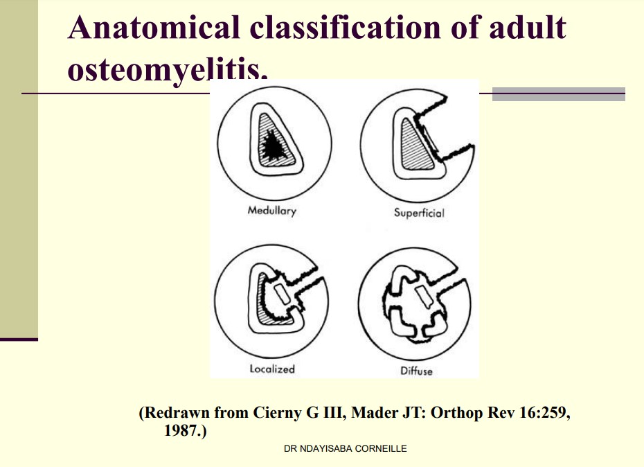
This Understanding Osteomyelitis note presented a comprehensive overview of osteomyelitis, detailing its pathophysiology, different types, and diagnostic measures. He elucidated the mechanism of haematogenous seeding, where pressure can cause an exudate or abscess to extend into the bone’s subperiosteal region, medullary cavity, or epiphysis. He also outlined four types of osteomyelitis: endosteal or medullary lesion, superficial osteomyelitis, localized infection with full-thickness cortical sequestration, and diffuse osteomyelitis lesions that are mechanically unstable.
Dr Corneille discussed various diagnostic tools, including radiography, radionuclide imaging, computerized tomography, sonography, and magnetic resonance imaging. Chronic osteomyelitis was characterized as an infected dead bone within compromised soft tissue, surrounded by sclerotic, relatively avascular bone. He also described a specific form of chronic osteomyelitis, Brodie’s abscess, which occurs most often in the lower extremities of young adults and is typically caused by low-virulence organisms.
He further examined sclerosing osteomyelitis, where the bone is thickened and sclerotic, but abscesses and sequestra are absent. He underscored its possible cause as an infection triggered by low-grade, possibly anaerobic bacteria. Towards the end of the presentation, Dr Corneille explained the characteristic ultrasonic features of acute osteomyelitis and how to interpret a three-phase bone scan to differentiate between osteomyelitis, cellulitis, and a septic joint.
Excerpt:
Understanding Osteomyelitis
Synopsis
Radiography
Radionuclide Imaging
Computerised Tomography
Sonography
Magnetic Resonance Imaging
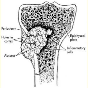
Pathophysiology of haematogenous seeding
When under pressure, exudate or abscess can extend through Volkmann canals into the subperiosteal region and from there into the medullary cavity or epiphysis.
Understanding Osteomyelitis
Anatomical criteria – consist of four types:
type I, an endosteal or medullary lesion;
type II, superficial osteomyelitis, limited to the surface of the bone;
type III- localized infection involving a stable, well-demarcated lesion characterized by full-thickness cortical sequestration and cavitation
type IV, diffuse osteomyelitis lesions that are mechanically unstable, either at presentation or after appropriate treatment
Reviews